Figure 5.

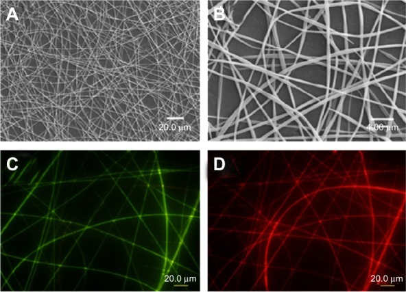
SEM and fluorescence images of the bicompartmental nanofibers composed of poly(NIPAM-co-SA) and PEGDMA compartments.
Notes: (A and B) SEM and (C and D) fluorescence images of the bicompartmental nanofibers composed of poly(NIPAM-co-SA) and PEGDMA compartments with separate fluorescence signals of (C) fluorescein and (D) Nile red channels in the dry state. Discrete nanofibrous structures with two fluorescence signals of the fluorescein and Nile red channels were observed. Scale bars are 20.0 μm in (A, C, and D) and 4.0 μm in (B).
Abbreviations: SEM, scanning electron microscopy; poly(NIPAM-co-SA), poly(N-isopropylacrylamide-co-stearyl acrylate); PEGDMA, polyethylene glycol dimethacrylates.
