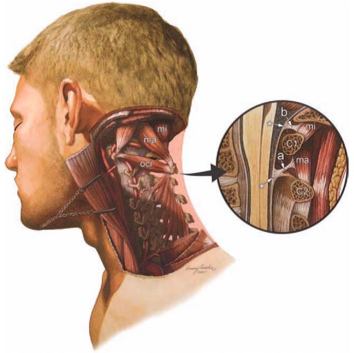
Figure 1.
Illustration of a dissection of the deep suboccipital region of the cervical spine. The rectus capitis posterior minor (RCPmi), rectus capitis posterior major (RCPma), and the obliquis capitis inferior (OCI) muscle fascia have communications with the dura mater via soft tissue. The encircled illustration (right) depicts a midsagittal dissection revealing the RCPma, RCPmi, and OCI muscles. The cervical myodural bridge (a) traverses the epidural space between the posterior elements of the C1 and C2 vertebrae. Both myodural structures link the suboccipital muscle fascia in to the cervical dura mater (*).Used with permission from: Magnetic resonance imaging investigation of the atlanto-axial interspace. Clin Anat. 2013 May; 26(4):444–9. Scali et al. (Original anatomical artwork by Frank Scali, D.C., and Danny Quirk)

