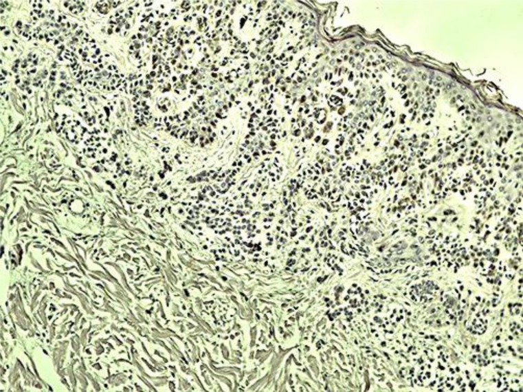
Fig. 2.
Mild hyperkeratosis and focal epidermal parakeratosis, mild spongiosis, extensive intracellular edema and focal lymphocytic exocytosis, moderate inflammatory infiltrate from the lymphocytes and histiocytes in his upper dermis and few polymorphonuclear cells perivascularly. Immunohistochemistry showed an intracellular accumulation of HHV-6 (brown stain).

