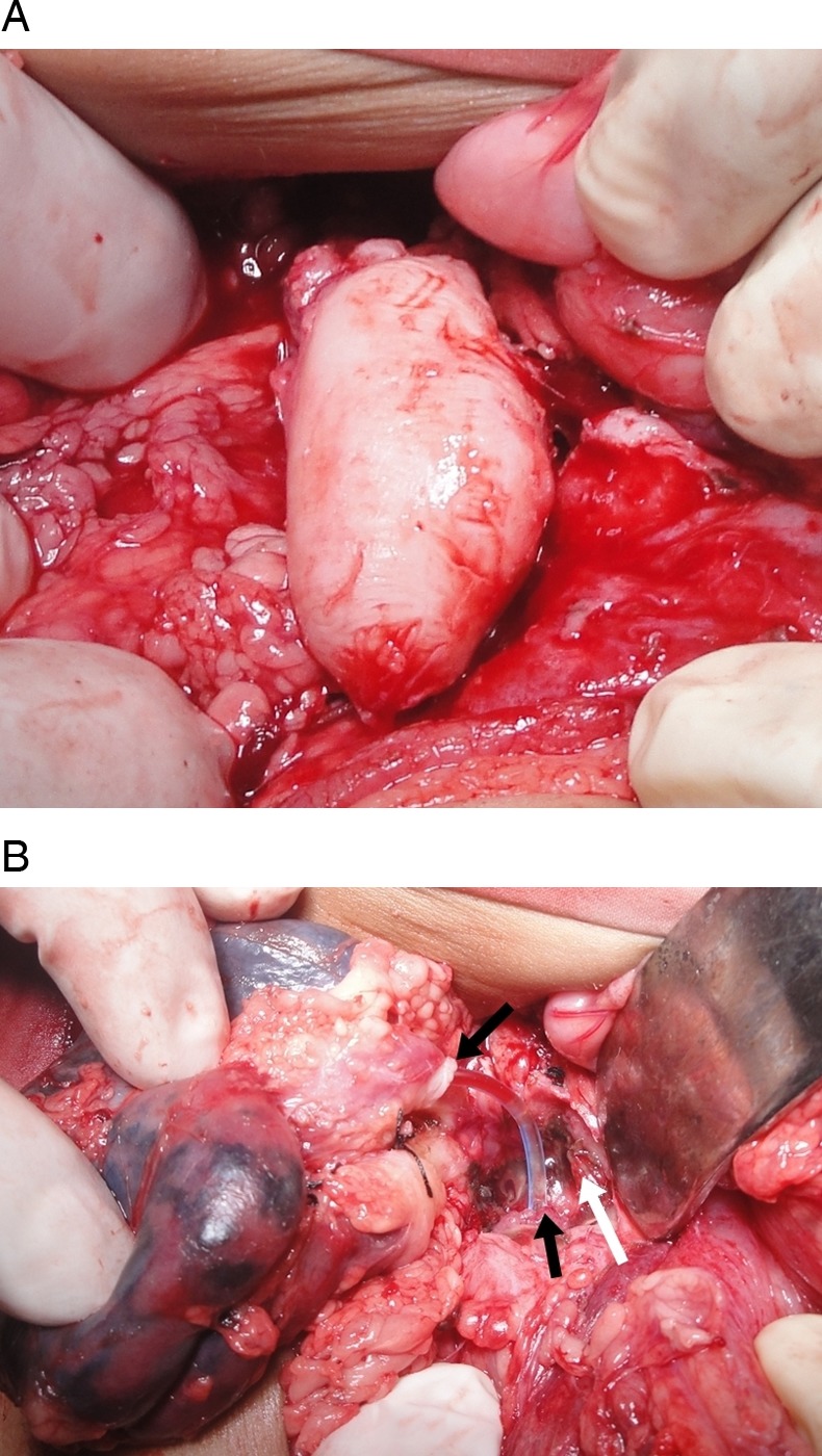
Figure 3.

Intraoperative image showing (A) a spherical, isolated, thick-walled cyst, (B) the renal pelvis, ureter and cyst opening into a common cavity; black arrows showing continuity of pelviureter where the feeding tube is passed; white arrow showing opening of the cyst into the cavity.
