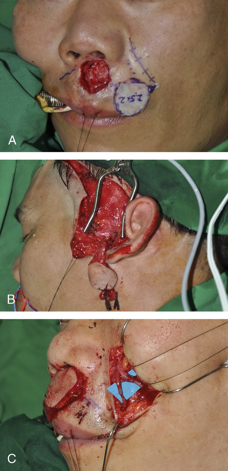
FIGURE 2.

The excision of deformed tissue creates a wound defect on the upper lip. This photograph shows the wound defect and its pattern (A). A hairy preauricular skin flap was harvested with a pedicle of superficial temporal vessels (B). A preauricular flap was inserted into the wound defect, and the flap pedicle was located in the subcutaneous tunnel. Anastomotic vessels of the nasolabial wound show a superficial temporal artery and a facial artery on the medial side, and a superficial temporal vein and a facial vein on the lateral side (C).
