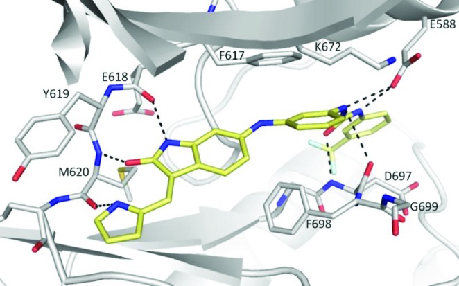
Figure 1.

X-ray crystal structure of compound 20 (stick representation; carbon in yellow, oxygen in red, nitrogen in blue, and fluorine in light blue) binding to the active site of TRKC kinase domain. The kinase domain is depicted as a cartoon with carbon atoms in gray, nitrogen in blue, and oxygen in red. Residues of the hinge region, the DFG motif, K672, and E588 are shown as sticks. Hydrogen bonds are shown as dashed lines.
