
Figure 3.
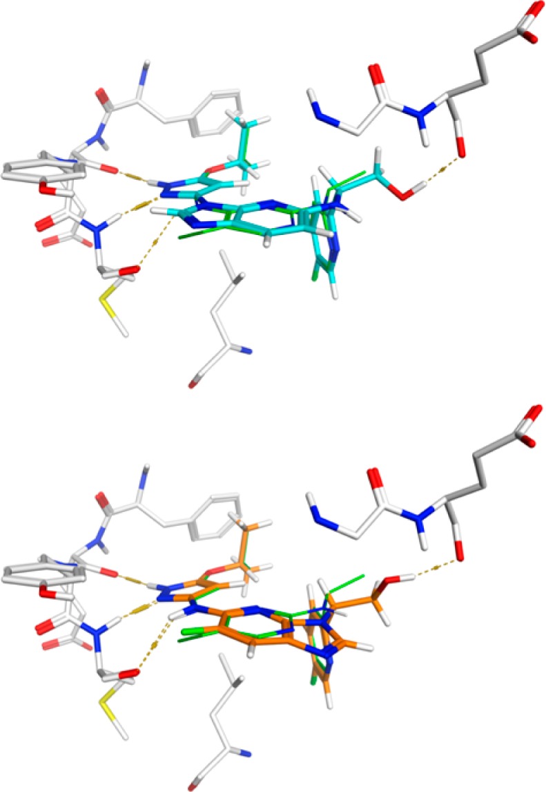
Proposed binding modes for 2d (top, cyan) and 3a (bottom, orange) in the TrkA binding site as they overlay with 1a (green). Hydrogen bonds are illustrated with dashed lines. The hydroxymethyl substituents in each example form an additional hydrogen bond with the backbone carbonyl oxygen of Glu 518 of the glycine-rich P loop.