Figure 3.

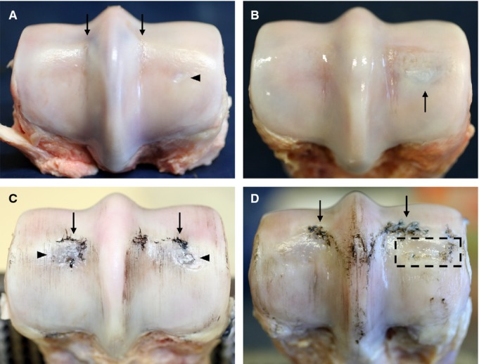
Macroscopic images of the distal end of the MC3 from younger (A,B) and older (C,D) animals. Lateral is to the left in all images. (A) Horse 5 showing an intact cartilage surface except for very slight linear fissures in the parasagittal grooves (see arrows). Note the small oval-shaped area of opaque white cartilage visible in the centre of the medial condyle region (see arrowhead). (B) Horse 4 with a slightly darkened region beneath undisrupted cartilage (see arrow). (C) Horse 14 showing minor wear lines, significant disruption at the transverse ridges (see arrows), and some darkening beneath the cartilage in the central condylar regions (see arrowheads), especially laterally. (D) Horse 13 showing a further example of cartilage disruption at the transverse ridge (see arrows). There is an area of the mid-condylar region where the joint surface is noticeably sunken in i.e. collapse of the articular surface (boxed region).
