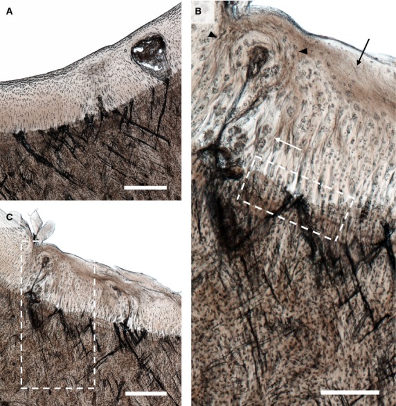
Figure 5.

Severe lesions in the hyaline cartilage and extensive microdamage in (A) the lateral and (B) the medial parasagittal groove of Horse 13. Note the high density of microcracks aligned in a cross-hatch configuration, with many large cracks extending across the calcified cartilage layer. Scale bars: 500 μm. (C) Enlarged image of the boxed region in (B). A number of changes can be seen in the hyaline cartilage including cell clustering (white arrow), regions of acellularity (black arrow), and regions of altered chondrocyte alignment suggestive of matrix remodelling (see black arrow heads). Cracks of various sizes can be seen in the calcified cartilage and subchondral bone. Scale bar: 250 μm.
