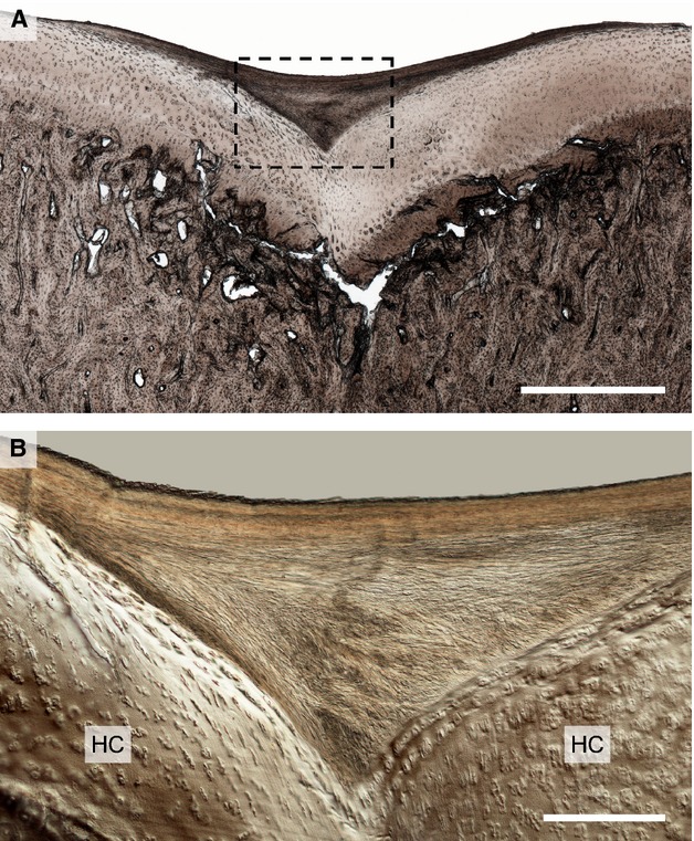
Figure 7.

Moderate damage in the mid-condylar region of Horse 5 showing the example of the first observed lesion type in a younger animal. (A) Low magnification view showing subchondral bone collapse and cartilage infolding in the mid-condylar region. Note the contrasting layer of ‘neo-tissue’ at the articular surface. Scale bar: 1 mm. (B) Higher magnification DIC image of the boxed region in (B). The neo-tissue that has formed is more fibrous in appearance and devoid of chondrocytes, thus being easily distinguished from the hyaline cartilage (HC) beneath, with which it is structurally integrated. Scale bar: 250 μm.
