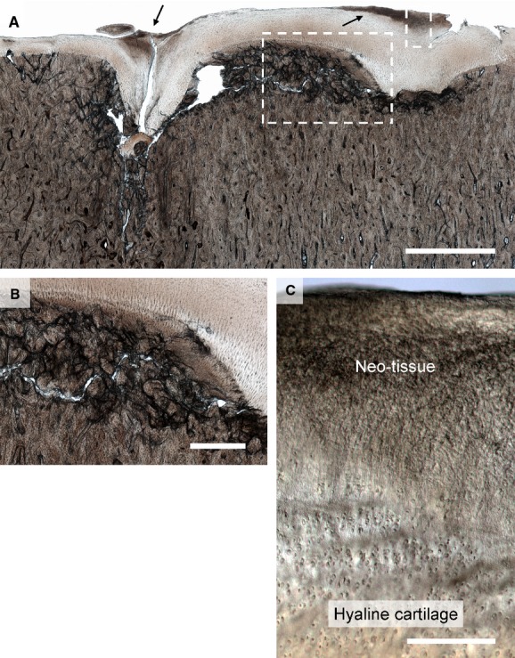
Figure 8.

Severe damage in the mid-condylar region of Horse 13. (A) Low magnification image. Subchondral bone collapse can be seen on the left with cartilage infolding. On the extreme right of the image there is evidence of what appears to be earlier subchondral collapse that may have stabilised. In both cases there is a greater amount of cartilage present than would be expected in a healthy region of the joint, with darker regions of neo-tissue at the articular surface (see arrows). The subchondral bone between the two collapsed regions has a comminuted appearance. Scale bar: 2.5 mm. (B) Enlarged view of left hand boxed region in (A) showing severe damage in the subchondral bone, which as a result has a comminuted appearance. Scale bar: 750 μm. Enlarged view of right hand boxed region in (A) showing the fibrous neo-tissue blending into the underlying hyaline cartilage. Scale bar: 200 μm.
