Figure 3.

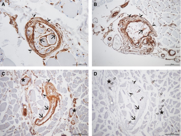
Muscle spindles of of different cadavers aged 71 (A), 97 (B), and 89 (C and D) years (A, cadaver no. 2 and B, cadaver no. 3, both vastus medialis; C and D, cadaver no. 4, bulbospongiosus), stained with antibodies against cav-1 (A-C) and neurofilament (D). Note the intense staining for cav-1 of all cellular layers of the muscle spindle sheath (arrowheads), regardless of the collagen content (‘normal’ appearance in A, thickened appearance in B). The sheaths around the individual intrafusal muscle fibres (the inner capsule) are intensely stained (black arrows in A and B). The perineural sheath of individual nerve bundles is intensely stained for cav-1 (stars in C and D). The sheath of the nerve fibres supplying the muscle spindle (black arrows in C and D) is continuous with the sheath of the muscle spindle (arrowheads). Small white arrows indicate single neurofilament positive nerve fibres. Scale bars: 50 μm (A), 100 μm (B–D).
