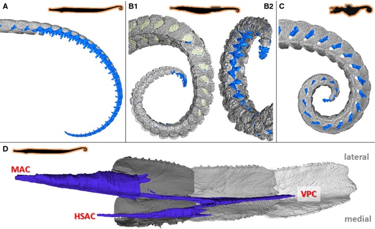
Figure 5.

Musculoskeletal organization in pipehorse. High resolution 3D reconstructions of CT-data on the caudal part of the tail in pipehorses (horizontal body posture, prehensile tail lacking a tail fin), represented by (A,D) Syngnathoides biaculeatus (type II pipehorse), (B) Solegnathus hardwickii (type II pipehorse) and (C) Acentronura gracilissima (type I/pygmy pipehorse). (A-C) Reconstruction of the caudal skeletal part of the tail (dermal plates in grey, vertebrae in blue). (D) Reconstruction of the conical myoseptal organization (dermal plates in grey, myosepta in blue), with a main anterior cone (MAC), ventral posterior cone (VPC) and a secondary hypaxial cone (sHAC).
