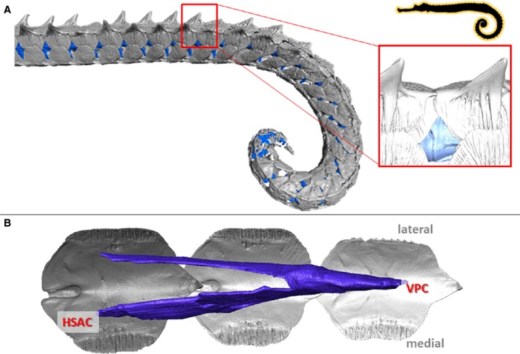
Figure 6.

Musculoskeletal organization in Haliichthys taeniophorus. High resolution 3D reconstructions of CT-data on the caudal part of the tail in H. taeniophorys (horizontal body posture, prehensile tail lacking a tail fin). (A) Reconstruction of the caudal skeletal part of the tail (dermal plates in grey, vertebrae in blue); detail shows a lateral view of the caudal spine on the dorsal plates fitting into an anterior groove on the subsequent plate. (B) Reconstruction of the conical myoseptal organization (dermal plates in grey, myosepta in blue), with ventral posterior cone (VPC) and a secondary hypaxial cone (sHAC).
