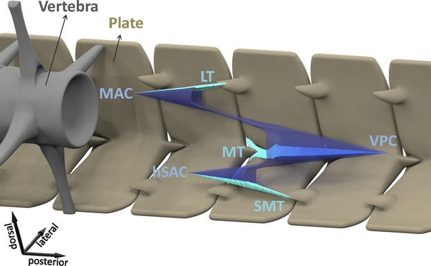
Figure 7.

Schematic overview of the conical organization of myosepta and tendons as found in the ancestral condition. Plates in brown, vertebra in grey, myosepta in blue (MAC, main anterior cone; VPC, ventral posterior cone; hSAC, hypaxial secondary anterior cone) and tendons in light green (LT, lateral tendon; MT, myorhabdoid tendon; SMT, secondary myorhabdoid tendon).
