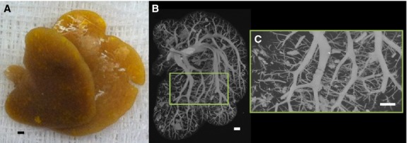
Figure 2.

(A) Photograph of only the caudate lobe after injection of yellow Microfil® contrast polymer into the hepatic vasculature. (B) Caudate lobe scanned in its entirety at 20 μm cubic voxel size. (C) Subvolume of caudate lobe scanned at high-resolution (5 μm cubic voxels). Scale bars: 1 mm.
