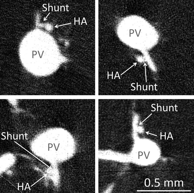
Figure 7.

Four examples of observed hepatic arteriolo-portal venular shunts in micro-CT images, observed by preparation method 1. These occur most frequently when a small portal vein branch comes off the main portal vein trunk close to the concomitant hepatic artery vessel.
