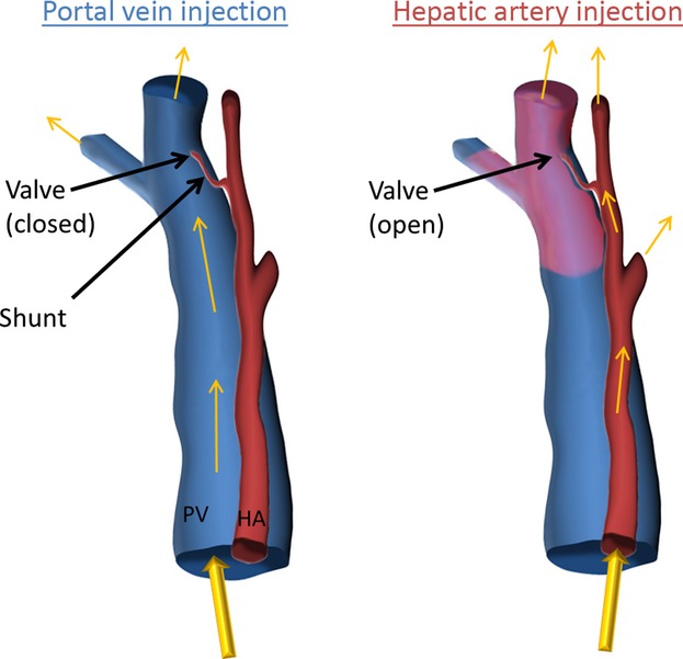
Figure 12.

Illustration of one-way valve-like mechanism of hepatic arteriolo-portal venular shunts. Where contrast is injected only into the portal vein, only the portal vein is present, since the shunt is closed and does not allow flow from the portal vein into the hepatic artery. Where contrast is injected only into the hepatic artery, the valve is opened (the portal vein being at low pressure due to no contrast being injected) and contrast fills both the hepatic artery and the portal vein. This illustration explains the results presented in Figs 4 and 5.
