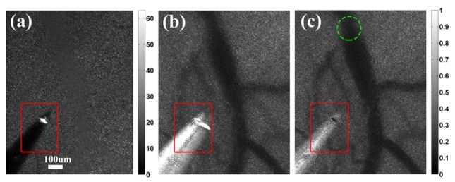
Fig. 2.

Raw speckle image and laser speckle contrast images in microscopic view. (a) Raw speckle image in microscopic view. The white area within the red square is the acupuncture needle tip, which was used as a reference for registration. (b) LSI image calculated without registration. (c) LSI image after registration. The green dashed circle indicates the focus of photothrombosis. It is clear that registration successfully removes motion artifacts and improves spatial resolution.
