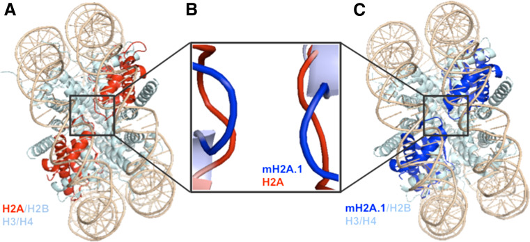
Fig. 3.
The L1 loops of mH2A.1 and canonical H2A are structurally distinct. a, c Crystal structure of nucleosomes containing canonical H2A (a, red) or mH2A.1 (c, blue) by in silico homology models. H2B, H3 and H4 are in light blue and DNA is in beige. b Superimposition of H2A and mH2A.1 LI loops illustrates a closer organization of the two mH2A.1 molecules compared to those of canonical H2A [160]

