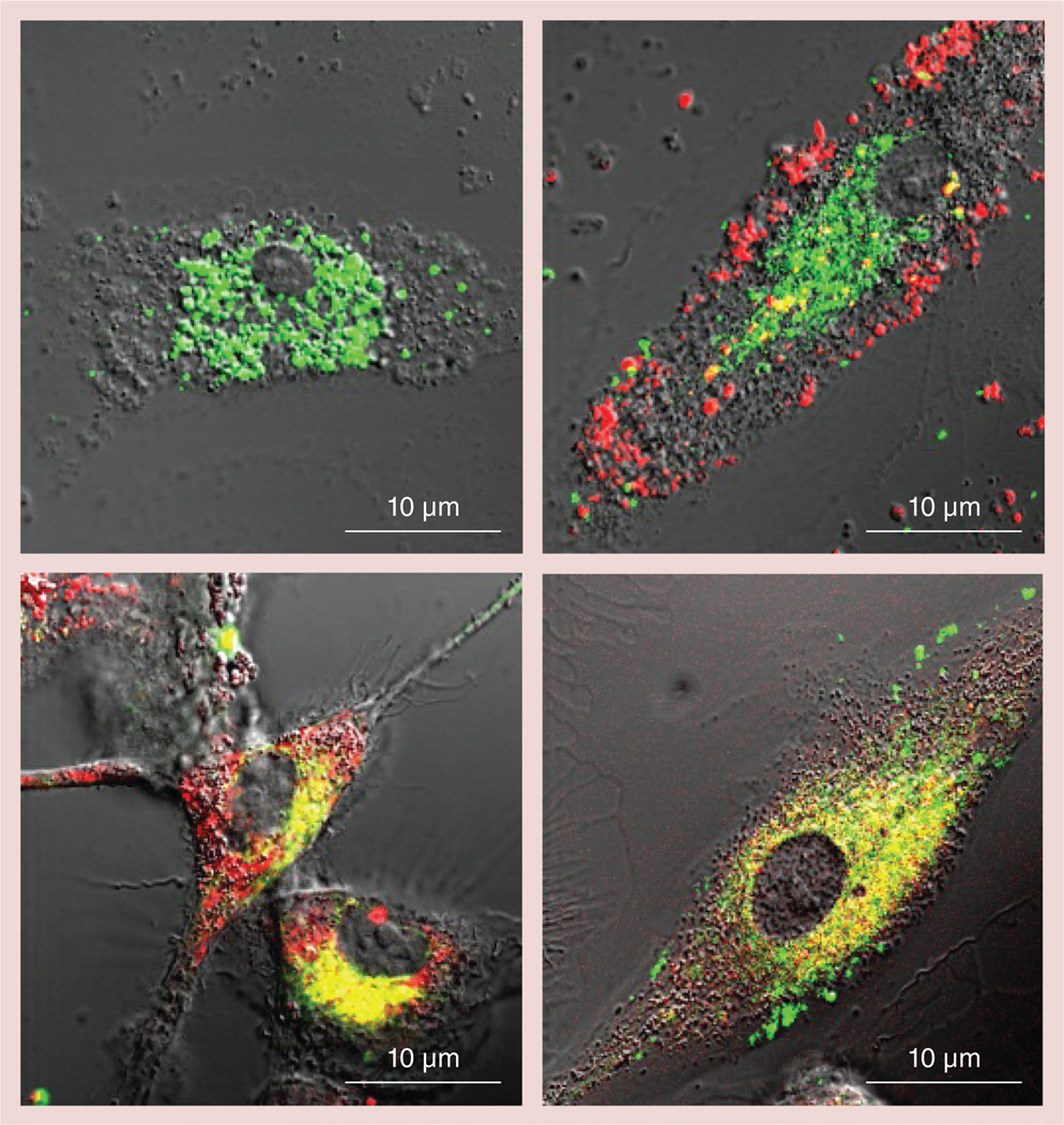
Figure 5. Intracellular distribution of catalase and block copolymer in bone marrow-derived macrophages.
Different nanozymes were prepared with Alexa Fluor® 488-labeled catalase (Invitrogen, CA, USA; green) and rhodamine isothiocyanate-labeled polymers (poly(ethyleneimine) [PEI] or PEI–polyethylene glycol [PEG]; red). Bone marrow-derived macrophages were incubated for 2 h with (A) catalase-alone, or different nanozyme formulations: (B) non-cross-linked (non-Cl) PEI/catalase; (C) non-Cl PEI–PEG/catalase (non-Cl-6); or (D) Cl PEI–PEG/ catalase (Cl-6-bis-(sulfosuccinimidyl)suberate sodium salt), washed with phosphate-buffered saline, and then visualized by confocal microscopy. (D) Stabilization of nanozyme structure by cross-linking resulted in practically total colocalization of the polymer and catalase inside macrophages. (C) PEG corona in non-Cl nanozyme partially prevented block ionomer complex dissociation. (B) The non-Cl block ionomer complexes with PEI polymer were completely dissociated.

