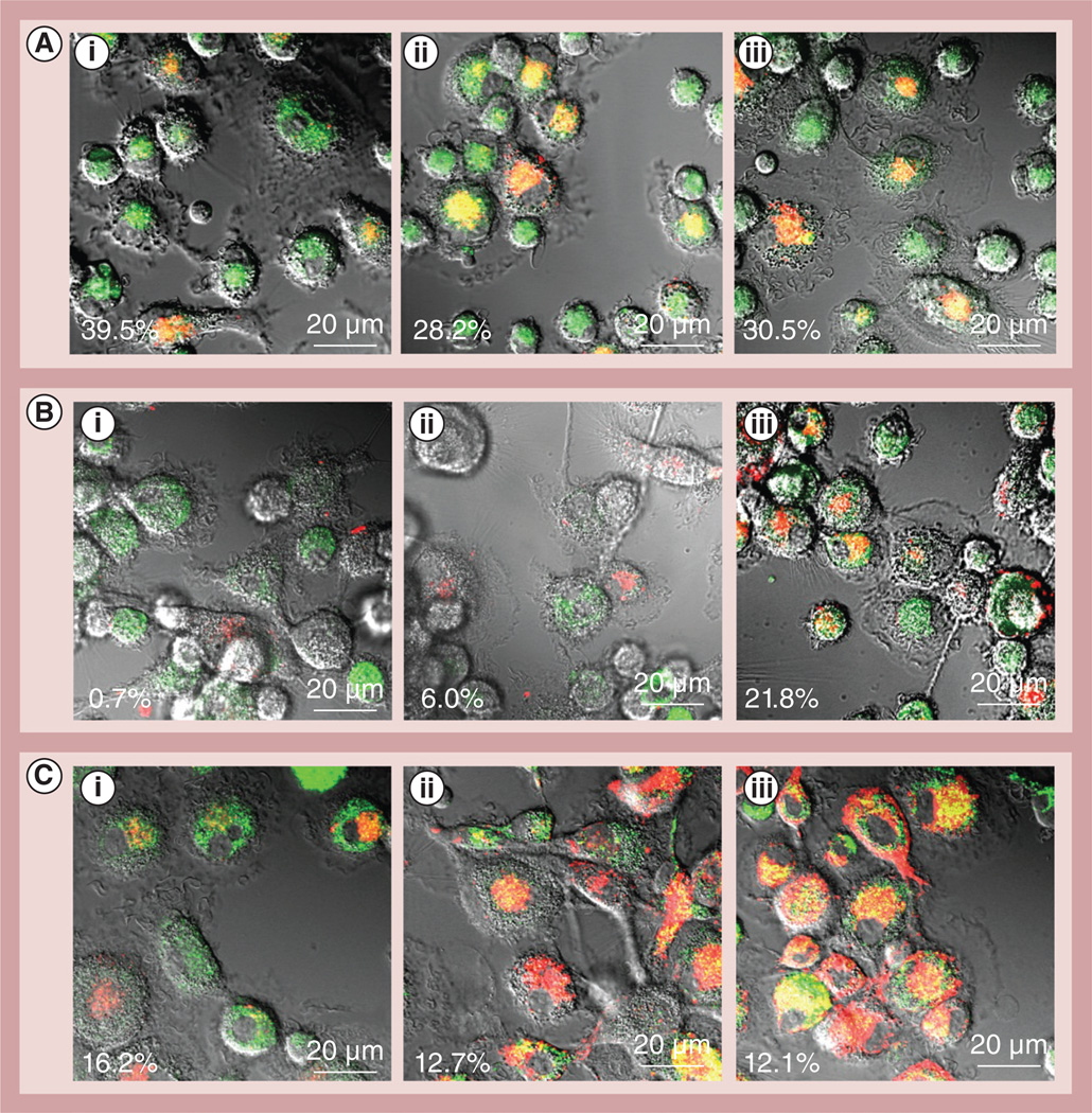
Figure 6. Nanozyme intracellular trafficking in macrophages.
Alexa Fluor® 680-labeled catalase (Invitrogen, CA, USA; red) was used to obtain cross-linked 6-bis-(sulfosuccinimidyl)suberate sodium salt. Live human monocyte-derived macrophages were incubated with cross-linked nanozyme for different times, and stained with: (A) LysoTracker® Green (Life Technologies, NY, USA; 150 nM), (B) ER-Tracker™ Green (Life Technologies; 1 µM), or (C) MitoTracker® Green (Life Technologies; 150 nM). The macrophages were incubated for (A,i, B,i & C,i) 5 min, (A,ii, B,ii & C,ii) 15 min and (A,iii, B,iii & C,iii) 30 min. Colocalization of nanozyme (red) and compartment staining (green) is manifested as yellow. Nanozyme accumulated first in acidified compartments and then reached mitochondria and endoplasmic reticulum. Refer also to Supplementary Table S3 for quantitative colocalization. The percentages in the bottom left hand of every image are the percentages of colocalization of nanozyme (red) and the compartment staining (green) specific for each slide, which is manifested in yellow colour.

