
Fig. 7.
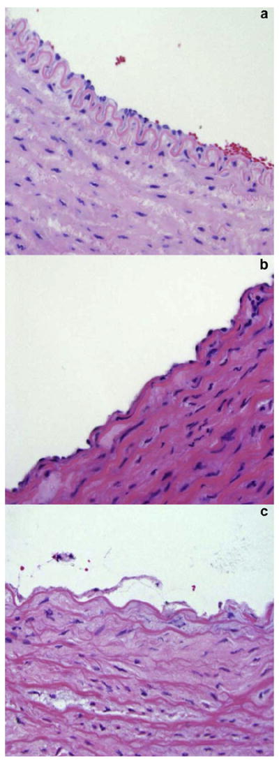
Representative microscopic images showing the endothelial and medial layers from carotid arteries of this study. After placement in the ex vivo flow system, most arteries showed overall normal aspect (a) with some areas of endothelial cell loss (b). Approximately 10% of the arterial rings showed signs of more severe distress, such as endothelial desquamation and medial edema (c).