
Figure 1. Identification of the RNA-binding region of JARID2.
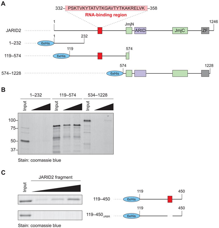
(A) Domain organization of human JARID2 and scheme of the 6xHis-fused truncations utilized in the mapping experiments.
(B) In vitro streptavidin pull-down after incubation of increasing concentrations of the indicated JARID2 recombinant fragments with biotinylated HOTAIR1–333. Input, 2 μg; titration, 2 and 4 μg.
(C) High resolution mapping of the residues of JARID2 necessary for RNA binding in vitro. The two indicated fragments (right) were incubated with HOTAIR1–333 and assayed as in (B). Input, 2 μg; titration, 1, 2, and 4 μg.
See also Figure S1.