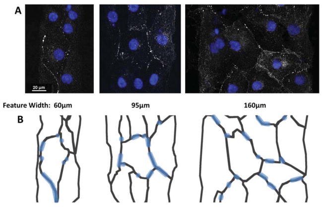
Fig. 6.
Connexin 43 expression of pure hESC-CMs seeded onto micropatterned matrigel/FN features of varying widths. Connexin 43 immunostaining with DAPI (A) shows a highly punctate and variable expression of gap junction plaques within the aggregates. Gap junction plaques were present along both lateral and axial edges of cardiomyocytes. A diagram (B) is included to indicate cell boundaries, as determined by phalloidin co-stain.

