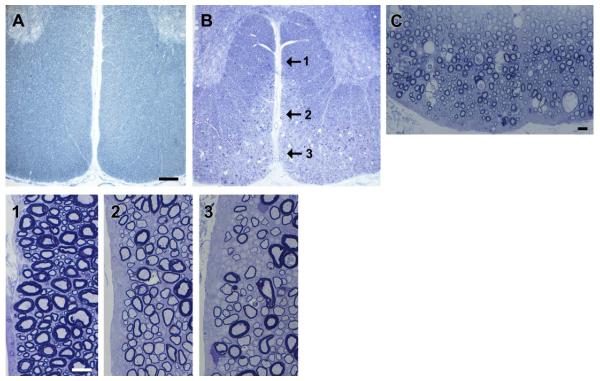
FIGURE 4.
Myelin vacuolation develops with time and axons remain non-myelinated. (A) The ventral column of a control mature dog shows even myelination throughout the white matter compared with an 11-month-old Chow that has a peripheral zone of abnormality of the superficial tracts (B). On higher power (C) many myelin vacuoles are seen close to the pia. Three differing patterns of myelination are present in the ventral column (1, 2, 3 arrows) shown on higher power below. The deep white matter is normally myelinated (1) while an adjacent area (2) contains mainly hypomyelinated fibers, and closer to the ventral surface of the cord (3), many axons remain nonmyelinated. Scale bars, A and B = 0.5 mm, C = 20 μm, 1–3 = 20 μm. [Color figure can be viewed in the online issue, which is available at wileyonlinelibrary.com.]

