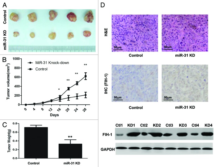
Figure 4. MiR-31/FIH-1 nexus controls the tumor growth of HCT116 xenografts in vivo. (A) Downexpression of miR-31 strikingly decreased the growth of HCT116 cells xenografted in nude mice. (B) The tumors were much bigger in control group than that in miR-31 knockdown group. (C) The tumors were much heavier in control group than that in miR-31 knockdown (miR-31KD) group. (D) Representative H&E-stained sections of the subcutaneous tumor tissues collected from control and miR-31KD groups were shown (up). FIH-1 expressions in the subcutaneous tumor tissues collected from control and miR-31KD groups were detected by IHC (middle, magnification of 200×) and western blotting (down). *P < 0.05, **P < 0.01.

An official website of the United States government
Here's how you know
Official websites use .gov
A
.gov website belongs to an official
government organization in the United States.
Secure .gov websites use HTTPS
A lock (
) or https:// means you've safely
connected to the .gov website. Share sensitive
information only on official, secure websites.
