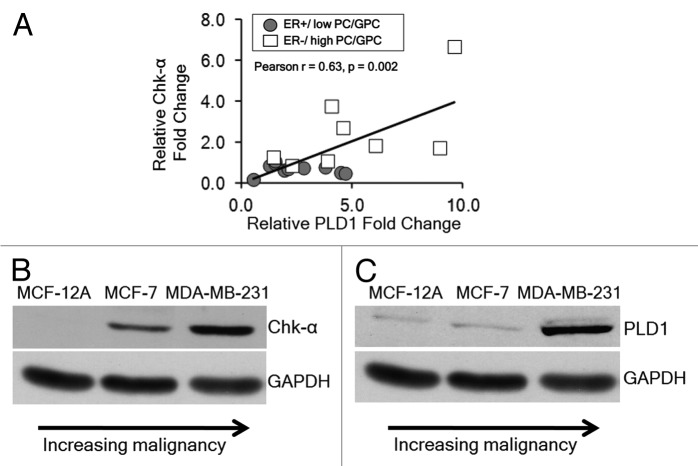
Figure 1. (A) Relative fold change in PLD1 mRNA and Chk-α mRNA in patient-derived tumor samples that are either ER+ (n = 11) or ER− (n = 8). (B) Immunoblots representing the protein expression of Chk-α in nonmalignant MCF-12A cells, non-metastatic ER+ MCF-7 cells and highly metastatic ER− MDA-MB-231 cells. (C) Immunoblots representing the protein expression of PLD1 in MCF-12A, MCF-7, and MDA-MB-231 cells. GAPDH was used as a loading control.

An official website of the United States government
Here's how you know
Official websites use .gov
A
.gov website belongs to an official
government organization in the United States.
Secure .gov websites use HTTPS
A lock (
) or https:// means you've safely
connected to the .gov website. Share sensitive
information only on official, secure websites.
