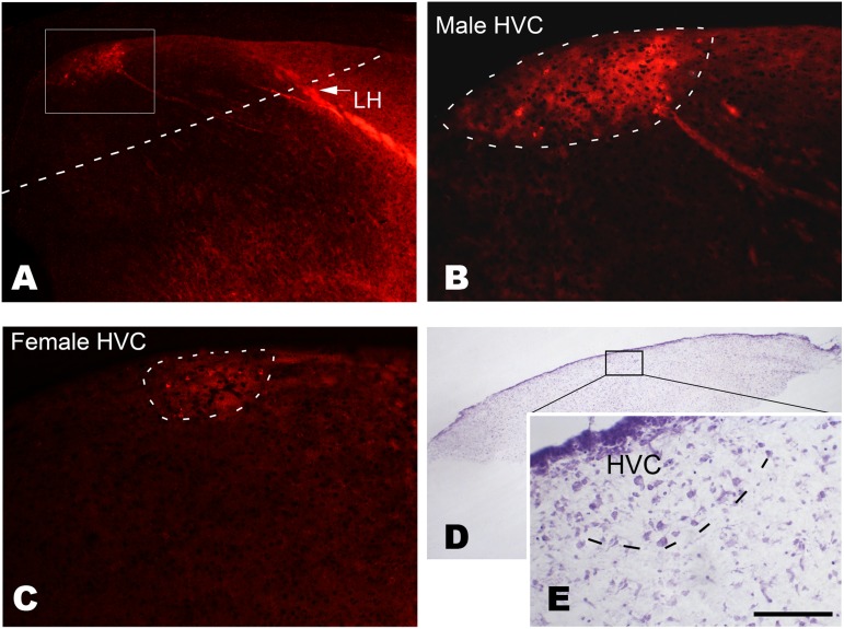
Figure 1. Determination of the location of the developing HVC at post-hatch day 15.
Following the injection of DiI into Area X, retrograde labeled cells are observed in the developing HVC of males (A and B) or females (C). The cells are located caudally to the lamina hyperstriatica (LH). D and E: A Nissl-stained cultured brain slice containing the developing HVC, which has many relatively larger cells compared with its surrounding areas (E). The cultured brain slice is obtained by cutting along the dashed line shown in A. The boxed area is amplified in B. Scale bar = 350 µm (A), 100 µm (B and C), 500 µm (D) and 75 µm (E).

