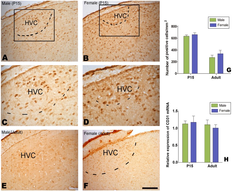
Figure 14. Distribution of Laminin-positive cells in HVC.
laminin-positive cells in HVC at 15 post-hatching days (P15, A–D) or at adulthood (E and F). Comparison of the number of laminin-positive cells at P15 and adult (G). H: Expression levels of CD31 mRNA relative to β-actin in the male and female HVC.

