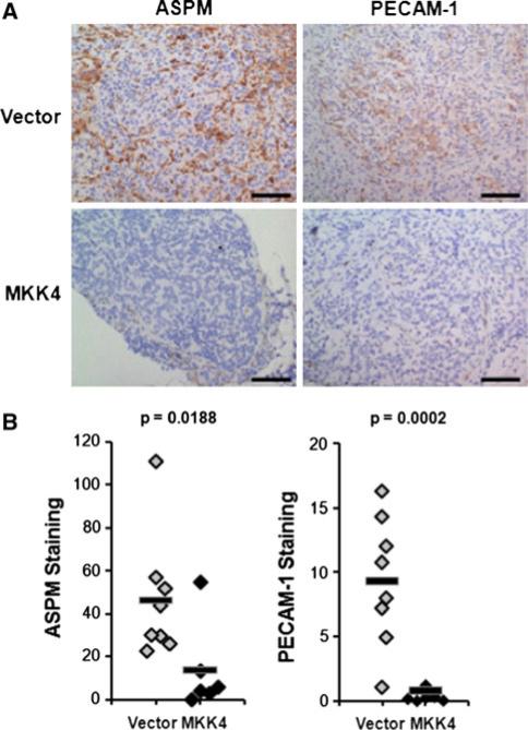
Fig. 4.
Early metastases expressing MKK4 have decreased ASPM and PECAM-1 staining in the cancer cells. Panel A IHC was performed on tissues from an independent cohort of 20 animals. Staining was performed as described in “Materials and methods” on frozen sections from tissues harvested 14 dpi. Representative images of ASPM and PECAM-1 staining in metastases formed by the two cell types are shown. Scale bars represent 100 μm. Panel B quantitative estimate of stain intensity using the ACIS Chromavision platform. The γ-axis was calculated by determining the average intensity of the brown pixels per unit area (IOD/10 mm2) for one sample from each animal. Differential protein expression was determined by comparing the staining intensity of MKK4-expressing metastases to metastases formed from cells expressing empty vector using a one-tailed t-test. Average staining intensity is indicated with a solid bar

