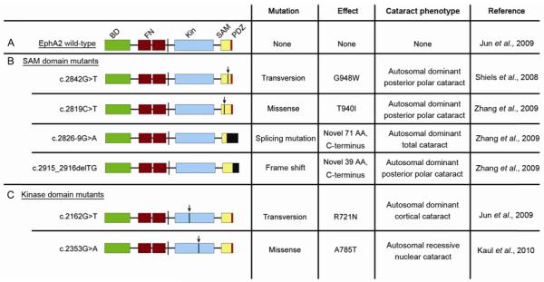
Figure 2.
Human EphA2 cataract mutations. A, Structure of wild-type EphA2. BD, ephrin ligand binding domain; FN, fibronectin repeats; Kin, kinase domain; SAM, sterile alpha motif; PDZ, PDZ domain. B, EphA2 SAM Domain cataract mutants. C, EphA2 kinase domain cataract mutants. Arrows denote relative location of point mutations in indicated mutations. Black boxes denote novel amino acid changes.

