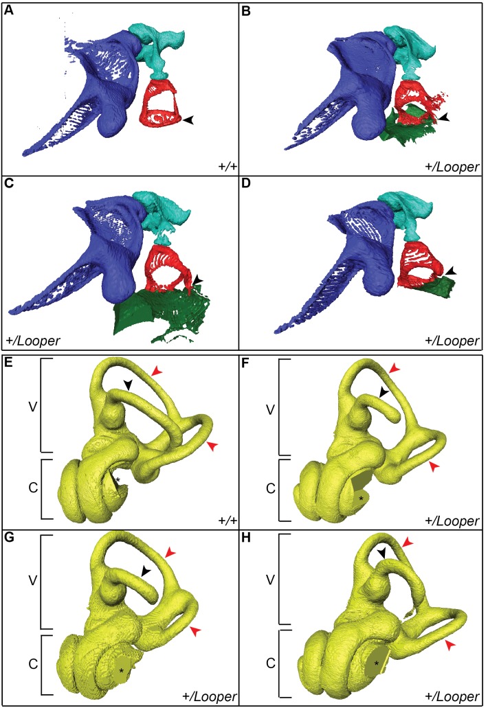
Figure 3. The Looper stapes is fused to the oval window and semicircular canals are abnormal.
µCT images of left middle (A–D) and inner (E–H) ears of Chd7+/+ and Chd7+/ Looper mice. A) Chd7+/+ and B–D) Chd7+/ Looper malleus (purple), incus (aqua) and stapes (red). The stapes footplate (arrowhead) is fused to the oval window of the cochlea (green) in Chd7+/ Looper mice. E) Chd7+/+ and F–H) Chd7+/ Looper vestibular apparatus (V) and cochlea (C). The lateral semicircular canal (black arrowhead) is incomplete and the posterior and anterior canals (red arrowheads) are hypoplastic in Cdh7+/ Looper mice. The missing section in each cochlea (*) was an artifact of the imaging/reconstruction process. n = 1 Chd7+/+ and 3 Chd7+/ Looper mice.

