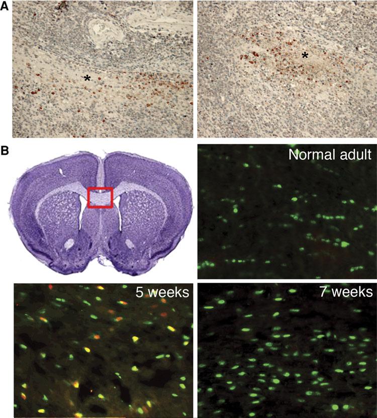
Fig 2.
Tcf7l2 protein is expressed in the brains of patients with PML and in the brains of the cuprizone-treated mice. a Brain sections of PML patients. Tcf7l2 protein (reddish brown) is expressed around the demyelination plaque (indicated by * symbol). The section is counter-stained with H&E. b Tcf7l2 protein (red) is expressed in the mouse corpus callosum (CC) after 5 weeks of cuprizone treatment. Before the treatment, only Olig2-positive cells (green) are seen in the CC region. After 5 week treatment, Tcf7l2 and Olig2 doublepositive cells (orange and yellow) are visible in the CC region. After 7 weeks, Tcf7l2 is no longer expressed, but Olig2-positive cells (green) remain in the CC region. The red frame indicates the region where the pictures are taken

