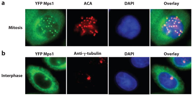
Figure 1.

Localization of Mps1 in vertebrate mitotic and interphase cells. (a) Kinetochore localization of yellow fluorescent protein (YFP) Mps1 in mitotic SW480 cells. Anti-centromere antibodies (ACA) and 4′,6-diamidino-2-phenylindole (DAPI) were used to stain kinetochores and chromosomes. (b) YFP Mps1 is localized to centrosomes and the cytosol during interphase. Centrosomes and nuclei were stained by anti-γ-tubulin and DAPI, respectively.
