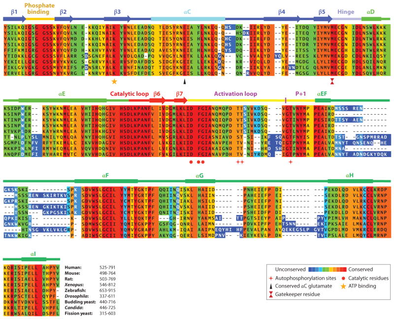
Figure 3.
Multiple sequence alignment of the kinase domain of Mps1 from representative species using PRALINE multiple sequence alignment (http://www.ibi.vu.nl/programs/pralinewww/). Amino acid conservation is shown by a color-coded heat map from unconserved to conserved. The secondary structure of Mps1 is shown above the Mps1. Key amino acid residues are shown below the alignment.

