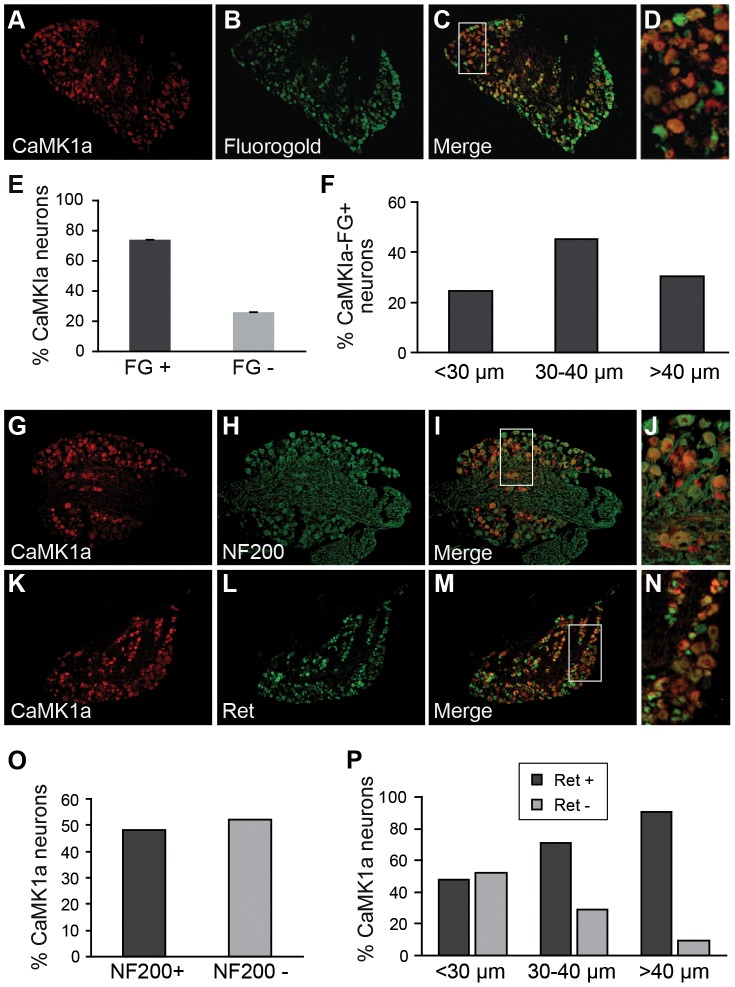
Figure 3. CaMK1a is preferentially induced in large diameter Ret+ neurons after axotomy.
(A–D). Combined CaMK1a immunohistochemistry and retrograde labelling with Fluorogold (FG) on L4–L5 DRG sections three days post-axotomy of the sciatic nerve. FG was applied at the cut nerve stump and specifically labels axotomized neurons. (E). Counts on DRG sections show that 74+/−2% of CaMK1a+ neurons are FG+. (F). Cell soma size distribution of CaMK1a+ neurons in DRG after sciatic nerve axotomy. (G–J). Double-immunofluorescent staining for CaMK1a and NF-200 on sections of L4–L5 DRG three days post-axotomy. (K–N). Double-immunofluorescent staining for CaMK1a and Ret on DRG sections three days post-axotomy shows numerous co-labelled neurons for both proteins. (O). Counts of CamK1a+NF200+ double-labeled cell reveals that about 50% of CaMK1a-positive neurons are NF200+. (P). Size repartition of CamK1a+Ret+ double-labeled cells showing that the vast majority of CaMK1a-positive neurons with medium-large cell soma diameter also express Ret.

