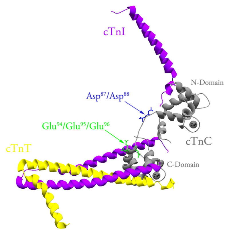
Figure 1. Location of the central helix residues within the structure of cTnC reconstituted into the cTn complex.

The figure shows a ribbon representation of the core domain of the cTn complex in the Ca2+ bound state (Protein Data Bank entry 1J1E (25)). CTnC, cTnI and cTnT are colored in grey, magenta and yellow, respectively. The Asp87/Asp88 residues (shown in blue) and Glu94/Glu95/Glu96 residues (shown in green) are located within the exposed middle segment of the central helix of cTnC. This figure was generated using Swiss-PdbViewer (56).
