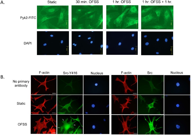
Figure 3. Pyk2 and Src accumulate in perinuclear and nuclear regions in response to OFSS.
(A) Immunofluorescence microscopy of osteoblast’s subjected to either static culture conditions or OFSS (30 minutes, 1 hour, or 1 hour+1 hour of rest). Slides were fixed immediately and processed for immunofluorescence using antibodies against Pyk2, followed by FITC-conjugated secondary antibodies. The nucleus was visualized using DAPI. Scale bars = 100 µm (B) OFSS induces accumulation of Src at perinuclear/nuclear regions in MLO-Y4 osteocytes. Immunofluorescence microscopy of MLO-Y4 osteocytes subjected to static culture conditions or OFSS for 20 minutes. Slides were fixed immediately and processed for immunofluorescence using antibodies against activated Src (Y416 followed by FITC-conjugated secondary antibodies). F-actin was visualized using Texas-Red Phalloidin and the nucleus was visualized using DAPI. White arrows indicate focal adhesions. Scale bars = 25 µm (B) Immunofluorescence microscopy of MLO-Y4 osteocytes subjected to static culture conditions or OFSS for 20 minutes. Slides were fixed immediately and processed for immunofluorescence using antibodies against total Src followed by FITC-conjugated secondary antibodies. F-actin was visualized using Texas-Red Phalloidin and the nucleus was visualized using DAPI. Scale bars = 25 µm.

