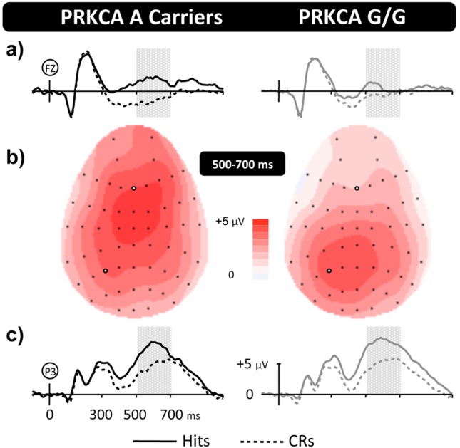
Figure 1. Distinct patterns of memory related brain activity for PRKCA polymorphisms.
Grand-average ERP old/new effects for PRKCA genotypes at representative Frontal (a) and Left-Parietal (c) electrodes, along with topographic maps (b) illustrating the distribution of old/new effects from 500–700 ms. The vertical scale indicates electrode amplitude (microvolts) and the horizontal scale change in time (milliseconds), with markers indicating the 500–700 ms window where significant results were found. The colour scale indicates Hit-CR difference size (microvolts). For both groups Hit ERPs are more positive going than CR from ∼300 ms post-stimulus onset (0 ms), reconverging by epoch end. Topographically dissociable maxima are evident across groups: parietally focused for G/G carriers and frontally focused for A carriers.

