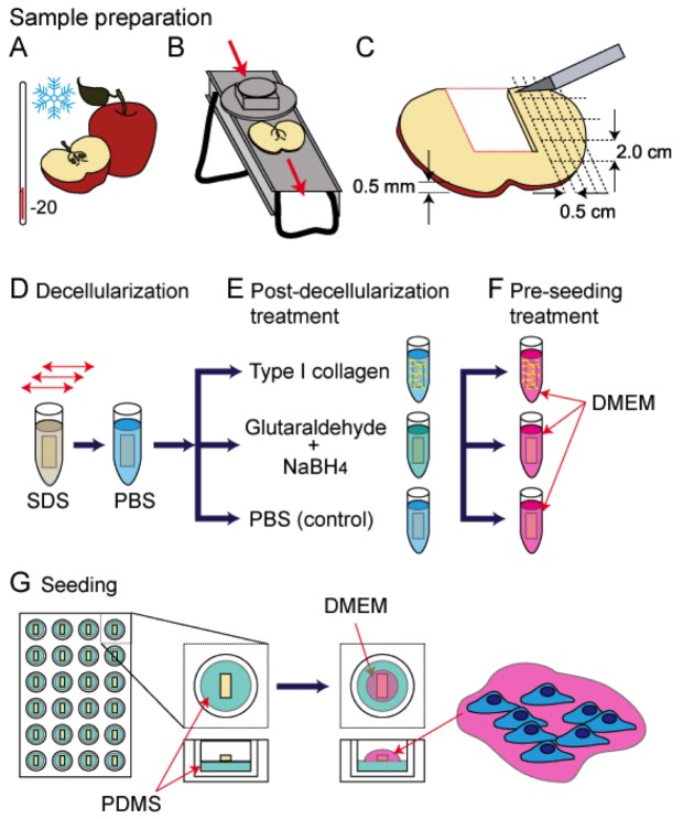
Figure 1. A cartoon schematic representing the apple tissue decellularization and mammalian cell seeding protocol used in this study.
A) McIntosh Red apples were exposed to −20°C temperatures for a max duration of 5 minutes, to increase the firmness of the outer apple hypanthium tissue. B) Uniform 1.2±0.1 mm thick slices of the apples were obtained using a mandolin slicer. Slices containing any of the ovary core of the apple were removed. C) The apple slices were cut into uniform 2.0 by 0.5 cm segments that were placed in individual microcentrifuge tubes. D) A 0.5% SDS solution was added to the microcentrifuge tubes and placed on a shaker for 12 hours at room temperature. The scaffolds were then rinsed repeatedly with PBS and allowed to incubate in a PBS solution with 1% streptomycin/penicillin and 1% amphotericin B for 6 hours at room temperature. E) The scaffolds were then coated with Type 1 collagen, chemically cross linked with glutaraldehyde or incubated in PBS. F) All the samples were then incubated in mammalian cell culture medium (DMEM) for 12 hours in a standard tissue culture incubator maintained at 37°C and 5% CO2. G) The scaffolds were placed in PDMS coated 24 well plates and a 40 µL cell suspension was placed on each. After 6 hours in the incubator the wells were filled with DMEM and cells cultured for up to 12 weeks.

