
Figure 2. Decellularized cellulose scaffolds.
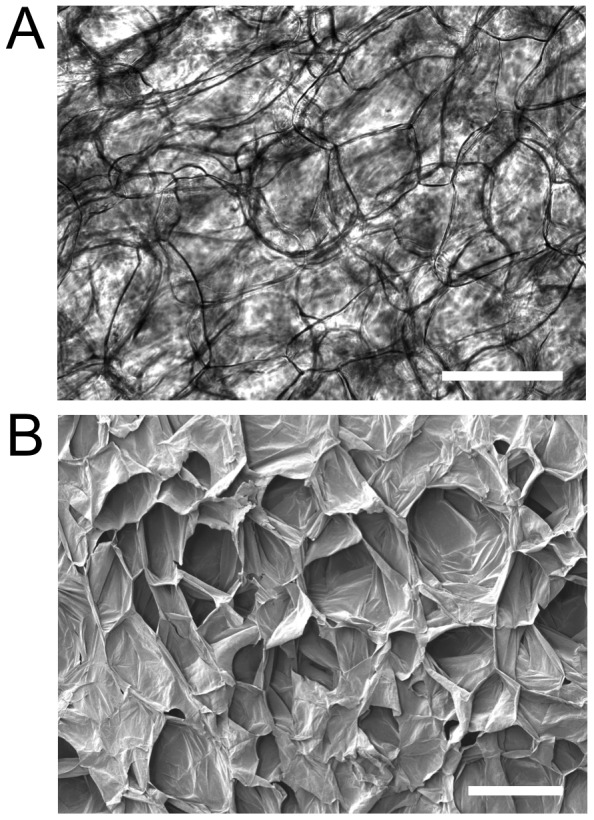
A) Phase contrast image of cellulose cell wall structure in a decellularized apple tissue sample. The dark lines correspond to distinct cellulose structures which form a three dimensional matrix. B) SEM image of a similar cellulose scaffold revealing its three dimensional nature and large cavities. Scale bar = 200 µm.