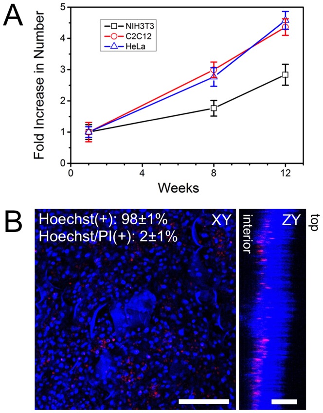
Figure 6. Cell proliferation and viability over time.
A) NIH3T3, C2C12 and HeLa cells were cultured individually in cellulose n = 3 scaffolds for 1, 8 and 2 weeks and then imaged with confocal microscopy after being stained with Hoechst 33342. Cells were counted at each time point and an increase in cell population is clearly observed. B) After 12 weeks of culture, C2C12 cells were fixed and stained with Hoechst 33342 (blue: viable cells) and Propidium iodide (PI) (red: apoptotic/necrotic cells). Confocal volumes were acquired and projected in the XY and ZY plane and reveal that cells have proliferated throughout the structure during the 12-week culture. The cells that are apoptotic/necrotic are found in deeper regions of the scaffold. The top and bottom surfaces of the scaffold are indicated. The number of live (Hoechst(+)) and dead (Hoechst/PI(+)) cells were counted and it was found that ∼98% of the cells within the scaffold are viable. Data is shown for C2C12 cells, but is similar for NIH3T3 and HeLa cells (data not shown). Scale bar: B = 200 µm for XY and 100 µm for ZY.

