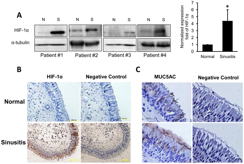
Figure 6. Sinus mucosa from chronic sinusitis patients shows high levels of HIF-1α and MUC5AC expression.
(A) The expression of HIF-1α in the control and sinus mucosa from four patients with sinusitis (western blot analysis). The increase of HIF-1α expression in sinusitis was 4.4-fold greater than in control tissue (n = 4; *p<0.05). (B) Immunohistochemistry with anti-HIF-1α antibody in normal (left) and sinusitis (right) tissues. High HIF-1α protein expression is indicated by the strong antibody reactivity in the epithelium of sinus mucosa from a sinusitis patient. (C) Immunohistochemistry with anti-MUC5AC antibody in normal (upper) and sinusitis (lower) tissues. Pronounced MUC5AC expression is seen in the epithelium from the sinusitis patient. Data is shown as mean ± standard deviation. (N; normal, S; sinusitis).

