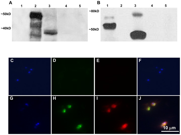
Figure 4. The gfp-bsd and MSA-1-Bm86ep transgenes are co-expressed in transfected parasites.
A–B Are the results from immunoblots using GFP (A) and Bm86 (B) specific antibodies. Lanes 1–5 represent recombinant MSA-1-BM86ep, recombinant GFP-BSD, whole cell lysate from the Tf-Bm86ep-gfp-bsd parasite line, whole cell lysate from Mo7 wild type, and non-infected RBC lysate control respectively. Molecular masses are indicated on the left of each panel. C–J Show results of fixed cell immunofluorescence using B. bovis Mo7 (C–F) and Tf-Bm86ep-gfp-bsd (G–J) infected erythrocytes from cultured cell lines. Parasites were stained with DAPI nucleic acid stain (C–G), GFP antibody labeled with Alexa Flour 488 (D–H), and Bm86ep antibody labeled with Alexa Flour 555 (E–I). Images were visualized using epifluorescence microscopy for each label, and a merged image was created (F–J). A 10 micron size bar is included in the bottom right panel.

