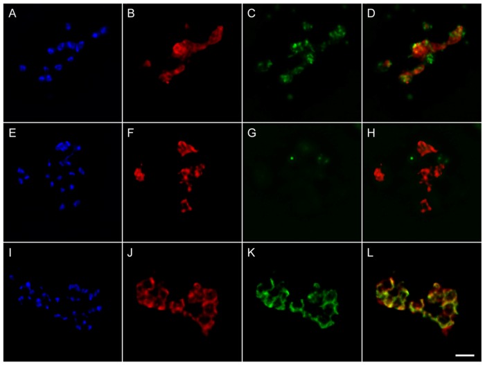
Figure 5. Bm86 epitopes are expressed on the surface of Tf-Bm86ep-gfp-bsd extraerythrocytic merozoites.
A–D represent permeabilized extraerythrocytic merozoites stained with DAPI (A), incubated with Bm86ep antibody labeled with Alexa Flour 647 (B), and GFP antibody labeled with Alexa Flour 488 (C). A merged image of panels B and C is shown in panel D. E–H represents identical staining procedures applied to non-permeabilized cells. I–L represent non-permeabilized extraerythrocytic merozoites stained with DAPI (I), and incubated with Bm86ep antibody labeled with Alexa Flour 647 (J), and the MSA-1 mAb Babb35 labeled with Alexa Flour 488 (K). A merged image of panels J and K is shown in panel L. A two micron size bar is included on the bottom right panel.

