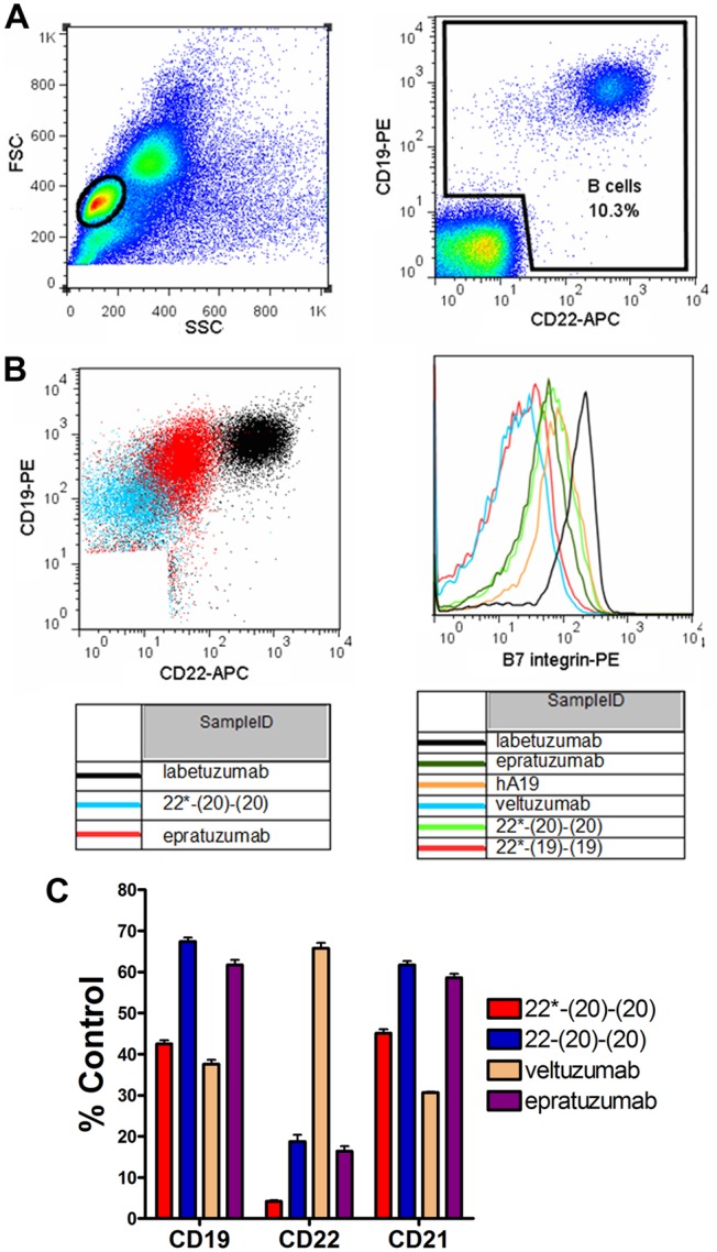
Figure 2. Analysis of trogocytosis by flow cytometry.

PBMCs were incubated overnight with 10 µg/mL of various mAbs or bsHexAbs prior to measurement of surface antigens by flow cytometry. (A) Gating of lymphocytes by forward vs. side scattering (Left) and B cells from the lymphocyte gate using CD19 and CD22 staining (Right) following treatment with control mAb (labetuzumab). (B) Example dot-plots comparing CD19 and CD22 staining on B cells following treatment of PBMCs with 22*-(20)-(20), epratuzumab and labetuzumab (Left) and histograms showing β7 integrin staining following treatment with the indicated mAbs or bsHexAbs (Right). (C) Trogocytosis mediated by Ck and CH3-based bsAbs. PBMCs were incubated overnight with 10 µg/mL 22*-(20)-(20), 22-(20)-(20), veltuzumab, epratuzumab or labetuzumab (control), prior to measurement of surface CD19, CD22 and CD21 by flow cytometry. Results are shown as the % MFI of the control treatment. Error bars, Std. Dev.
