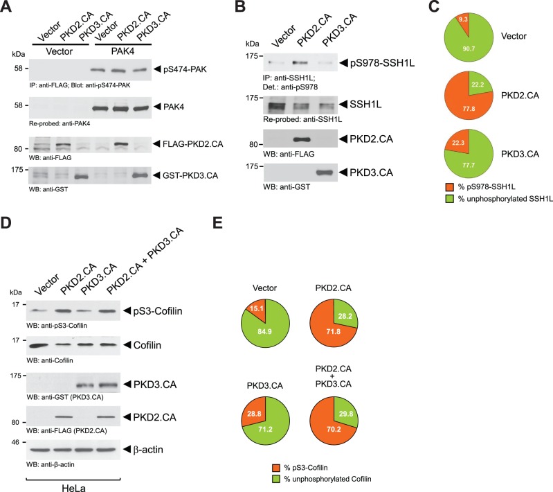
Figure 5. Increased PKD2 and PKD3 activities do not further enhance PAK4 activity, but inactivate SSH1L.
A: HeLa cells were co-transfected with vector control or FLAG-tagged PAK4 and FLAG-tagged constitutively-active PKD2 or GST-tagged constitutively-active PKD3, as indicated. Cells were lysed, PAK4 was immunoprecipitated (anti-FLAG), samples subjected to SDS-PAGE, transferred to nitrocellulose and analyzed for PAK4 activity by immunostaining with anti-pS474-PAK4 antibody. After stripping samples were re-probed with anti-FLAG (total PAK4). In addition, cell lysates were analyzed by Western blot (input control) for expression of PKD2.CA (anti-FLAG) and PKD3.CA (anti-GST). B: HeLa cells were transfected with vector control, FLAG-tagged constitutively-active PKD2 or GST-tagged constitutively-active PKD3, as indicated. Cells were lysed, endogenous SSH1L was immunoprecipitated (anti-SSH1L), samples subjected to SDS-PAGE, transferred to nitrocellulose and analyzed for SSH1L phosphorylation at S978 by immunostaining with anti-pS978-SSH1L antibody. After stripping samples were re-probed with anti-SSH1L (total SSH1L). In addition, cell lysates were analyzed by Western blot (input control) for expression of PKD2.CA (anti-FLAG) and PKD3.CA (anti-GST). C: Percentage of S978-phosphorylated and unphosphorylated SSH1L was calculated and depicted in a pie graph. D: HeLa cells were transfected with control vector, FLAG-tagged constitutively-active PKD2 or GST-tagged constitutively-active PKD3, as indicated. Amount of endogenous pS3-phosphorylated cofilin and total cofilin was determined by Western blot analysis (anti-pS3-cofilin and anti-cofilin for total cofilin). E: Percentage of S3-phosphorylated and unphosphorylated cofilin was calculated and depicted in a pie graph. Experiments shown in A, B and C were performed at least three times with similar results.

