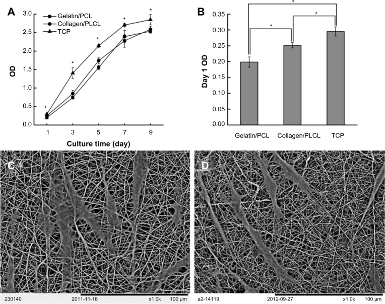
Figure 4.
Adherence and proliferation of HUSMCs on various materials.
Notes: SEM images of HUSMCs grown on gelatin/PCL (A) and collagen/PLCL (B) electrospun fibrous membranes for 1 day. Cell proliferation (C) and adhesion rate (D) on various materials measured by a CCK-8 kit.
Abbreviations: PCL, polycaprolactone; PLCL, poly(l-lactic acid-co-ε-caprolactone); SEM, scanning electron microscope; HUSMC, human umbilical arterial smooth muscle cell; CCK-8, Cell Counting Kit-8; TCP, tissue culture plate; OD, optical density.

