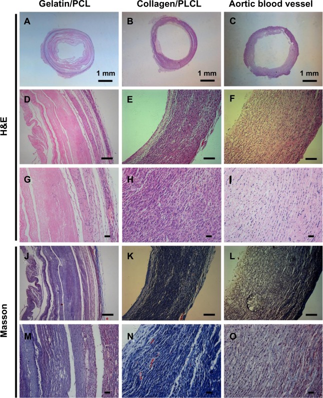
Figure 6.
Histological images of cell scaffold constructs with H&E and Masson’s trichrome staining at 6 weeks in vivo.
Notes: Panels A, D, G, J and M appear more heterogeneous with a large number of non-degraded scaffolds and less collagen fibers. Panels B, E, H, K and N show relatively homogenous vessel-like tissue structures with bands of collagen fibers formed. A newborn pig aortic blood vessel was used as a positive control (C, F, I, L and O). Scale bars: 100 μm.
Abbreviations: H&E, hematoxylin and eosin; PCL, polycaprolactone; PLCL, poly(l-lactic acid-co-ε-caprolactone).

